The following document was written by Mr Vik Veer MBBS(lond) MRCS(eng) DoHNS(eng) in Dec 2007. You may use the information here for personal use but if you intend to publish or present it, you must clearly credit the author and www.clinicaljunior.com
This site is not intended to be used by people who are not medically trained. Anyone using this site does so at their own risk and he/she assumes any and all liability. ALWAYS ASK YOUR SENIOR IF YOU ARE UNSURE ABOUT A PROCEDURE. NEVER CONDUCT A PROCEDURE YOU ARE UNSURE ABOUT.
QUINSY
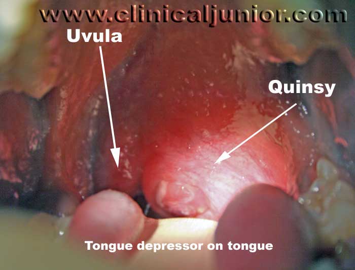
This is pus that has collected in the space between a tonsil and the wall of the mouth creating a large abscess (quinsy or peritonsillar abscess). The patient will be unable to open his/her mouth (trismus) the patient is very unlikely to have a quinsy without trismus as one of the signs. They will almost always have a altered ‘hot potato’ voice with a fever.
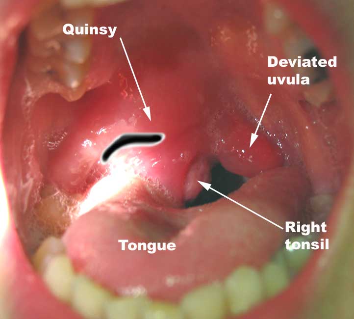
On examination there will be, and a large erythematous mass superior lateral to one of the tonsils. This mass will push the uvula over to the opposite side but rarely this is required to make the diagnosis.
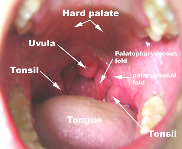
This first image is to show you what normal is - what it should look like...
 The image below shows a quinsy with the tongue and uvula shown as well. As you can see the uvula and the tonsil have been shifted over to the right to make way for the abscess.
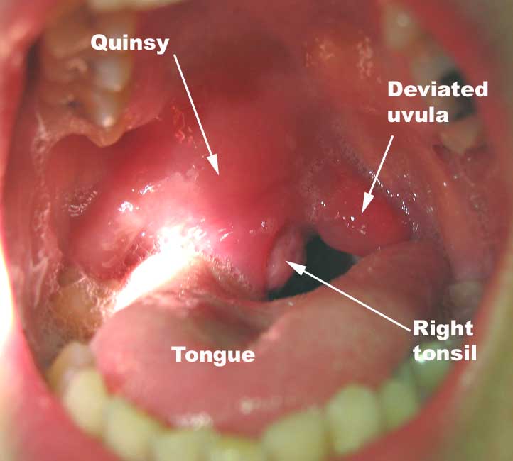
Here is another quinsy this time on the right side.
Management would be:
- IV cannula and take bloods – FBC, U&E’s, LFT’s, CRP, glandular fever screen
- Give iv Benzyl penicillin 1.2g QDS IV and metronidazole 500mg TDS IV
- Diclofenac 50mg orally TDS
- Codeine phosphate 60mg oral QDS
- Paracetamol 1g oral / IV
- Difflam sprays two sprays every 4 hours
- Fluid replace with adequate amounts of normal saline (normally a litre or more) if they look clinically dry
The abscess needs to be drained and depending on your hospital and time of admission this is either done by the ENT SHO oncall or admitted to the ward to be aspirated in the morning.
 Aspiration of a quinsy is potentially hazardous as the carotid artery is only a few centimetres from the tip of the needle. Don’t try this without first being guided by an experienced ENT doctor first.
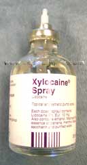
The technique is to spray the quinsy with some xylocaine spray and then inject the surface mucosa overlying the quinsy with 1% lignocaine with the smallest needle you can find (orange or dental needles).
It seems there are a lot of people who would not use local anaesthetic before aspirating or incising a quinsy. i've tried with and without and i am sure that it is much less painful for the patient using injected lignocaine rather than just sprayed LA. It does take more time (about 3 mins or so).
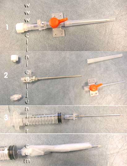
Whilst that is working get the largest cannula you can find (brown) and remove everything (plastic cannula, and other attachments) leaving only the needle, which can now be attached to a 20mls syringe.
Over this wrap sticky tape around the base of the needle until only the top 1.5 – 2cm of the tip of the needle is exposed. This will prevent you from going too deep with your needle. (see the picture steps on the right)
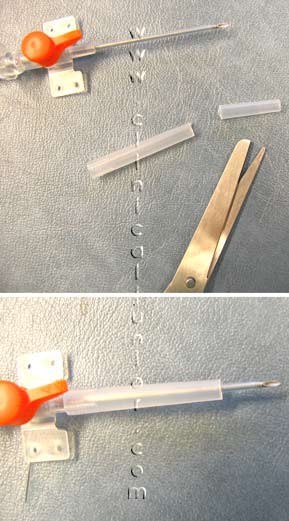
You can try a different method to make a safe quinsy aspiration kit - you can cut off the cannula protector at the appropriate level and leave it on so that you may use this to stop the needle from diving in too deep (see picture below right)
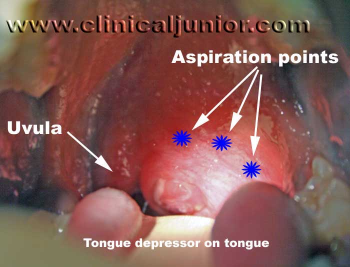
 Using a headlight and a tongue depressor, and steadying the patient insert the needle deep into the quinsy where it is pointing or looks largest, staying as horizontal and straight as possible. Classically you place the needle at the point where a vertical line up from the molar teeth and a horizontal line parallel to the base of the uvula meet. Every quinsy is subtly different and so you will improve your technique with experience.
The image below shows a quinsy with some aspiration points that should be a rough guide for you. Sadly it is very difficult to take pictures of a quinsy due to the trismus. Therefore also this picture doesn't give you a good idea of where excatly to aim - but i hope it gives you a start.
Normally one can expect about 5-10 mls of green / yellow pus to be aspirated. This should reduce the quinsy and allow the patient to open his/her mouth and improve his/her voice almost instantly.
If it hasn’t helped or you didn’t get any pus try 3 times in total and then move on to incision. Some people go straight to incision in the first instance but this is much more painful and requires more experience. In these cases it is likely the abscess has become loculated and is difficult to aspirate completely.
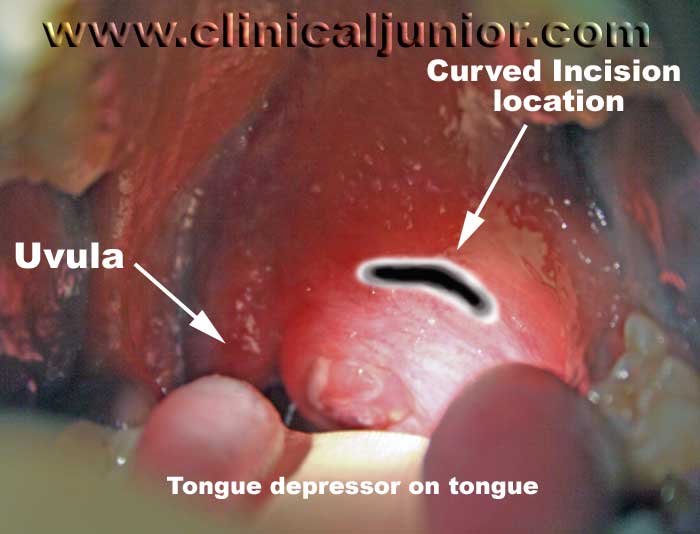
With the local anaesthetic still working and similar preparation make a curved incision in the same area as the aspirations and open up the abscess. I then use a spatula to break down the loculations within the abscess to make sure all the pus will drain appropriately. you should also use a tilly's or something similar to open up the cavity and allow the pus to drain more freely. This is very painful even with LA and should be done with a confident hand.
The image below shows a rough incision location - because the picture is off centre the incision looks more central than it really is. You should ask a senior if you are not sure where to incise.
Here is another example of where you should be incising to cure a quinsy. The original image is above before management.
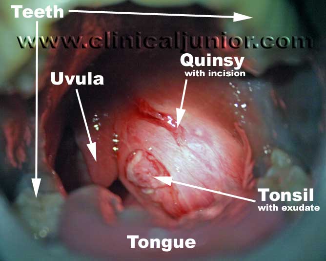
The picture below shows the above quinsy that has been incised and is half drained. In total about 12-13mls of pus was drained from this quinsy.
Patients stay in for 24 hours after this procedure as there is a risk of laryngospasm and of the abscess recollecting within the next 12 hours. Give the patient the medication mentioned above and admit.
Further Reading
EMEDICINE - Peritonsillar Abscess : Article by Ninfa Mehta
Peritonsillar Abscess (aka Quinsy) : Practical ENT For Primary Care Physicians web site
EMEDICINE - Tonsillitis and Peritonsillar Abscess : Article by Udayan K Shah